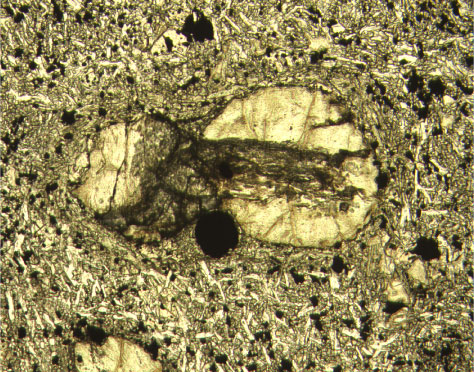
Figure F22. Photomicrograph of Cr spinel phenocryst (large dark spot) adjacent to olivine phenocryst (with Cr spinel inclusion) in Unit 17 (Sample 197-1206A-38R-1 [Piece 2B, 55-58 cm]) (plane-polarized light; field of view = 1.25 mm; photomicrograph 1206A-349).