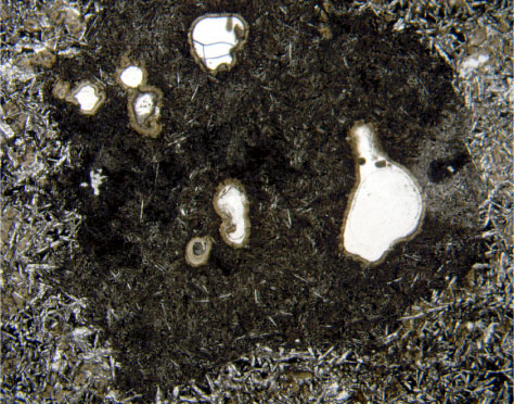
Figure F25.
Photomicrograph of segregated material surrounding vesicles in Unit 4 (Sample
197-1206A-8R-1 [Piece 1, 2-3 cm]
) (plane-polarized light; field of view = 5 mm; photomicrograph 1206A-316).