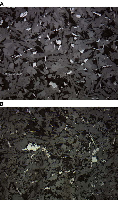
Figure F26. Photomicrographs showing skeletal octahedral and dendritic titanomagnetite morphologies (reflected light; field of view = 1.25 mm). A. Sample from Unit 1 with relict Cr spinel (lower, middle) (Sample 197-1206A-3R-2 [Piece 8, 94-96 cm]) (photomicrograph 1206A-291). B. Subunit 18b (Sample 197-1206A-41R-1 [Piece 5A, 22-24 cm]) (photomicrograph 1206A-366).