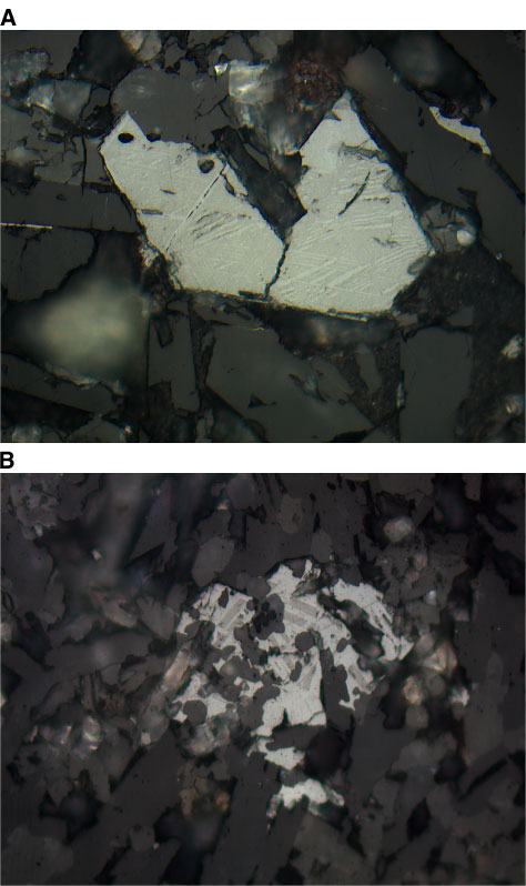
Figure F29. Photomicrographs showing the development of maghemite along cleavage planes and fractures (see also Fig. F28A) (reflected light; field of view = 0.25 mm). A. Unit 10 (Sample 197-1206A-24R-3 [Piece 2, 10-12 cm]) (photomicrograph 1206A-335). B. Unit 5 (Sample 197-1206A-15R-3 [Piece 15, 87-89 cm]) (photomicrograph 1206A-326).