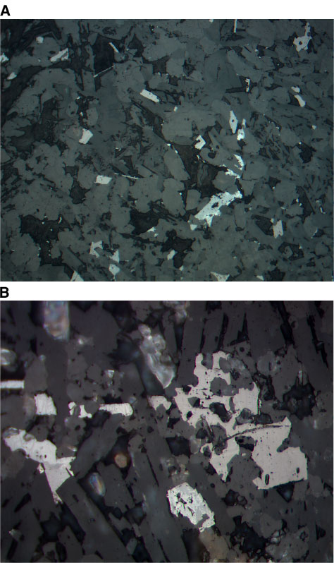
Figure F30. Photomicrographs showing the differential development of maghemite around groundmass titanomagnetite (reflected light). A. Unit 6 (Sample 197-1206A-18R-1 [Piece 4, 80-83 cm]) (field of view = 0.625 mm; photomicrograph 1206A-375). B. Unit 5 (Sample 197-1206A-15R-3 [Piece 15, 87-89 cm]) (field of view = 0.25 mm; photomicrograph 1206A-327).