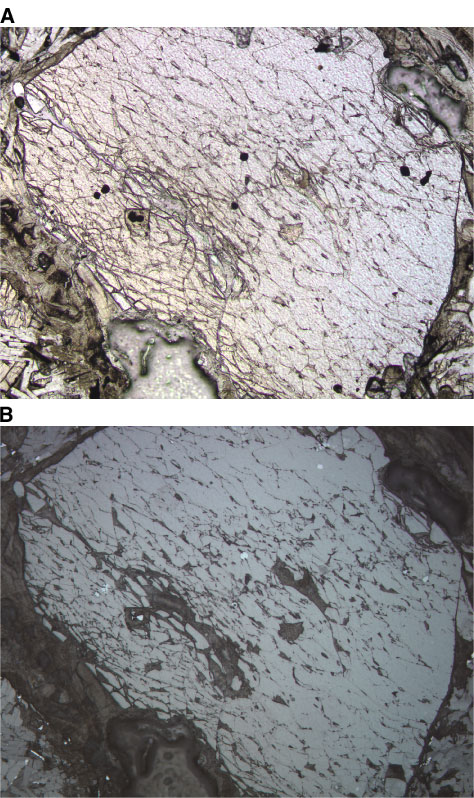
Figure F31. Photomicrographs showing Cr spinel inclusions (dark spots) in unaltered olivine phenocrysts in Unit 1 (Sample 197-1206A-4R-3 [Piece 4A, 72-74 cm]) (field of view = 1.25 mm). A. Plane-polarized light; photomicrograph 1206A-295. B. Reflected light; photomicrograph 1206A-296.