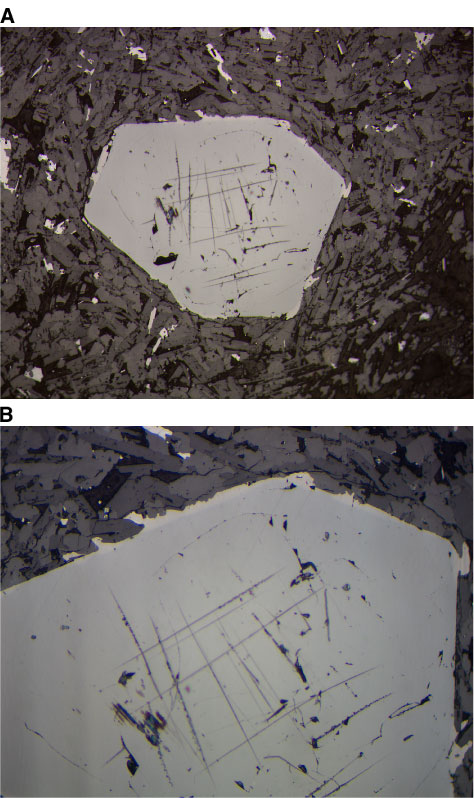
Figure F34. Photomicrographs of a discrete Cr spinel phenocryst in Unit 6 (Sample 197-1206A-18R-1 [Piece 4, 80-83 cm]) (reflected light). A. Euhedral morphology of the discrete Cr spinel phenocrysts (field of view = 1.25 mm; photomicrograph 1206A-379). B. Close-up of the view shown in A to emphasize the titanomagnetite overgrowth on the Cr spinel phenocryst, which also contains ilmenite oxidation lamellae (i.e., extreme right of the picture) (field of view = 0.625 mm; photomicrograph 1206A-380).