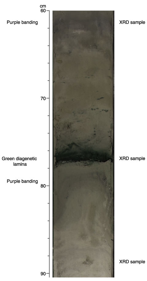
Figure F7. Close-up digital photograph showing a well-defined pale green lamina with diffuse purple banding both above and below (interval 198-1210B-3H-5, 60-90 cm). Locations of X-ray diffraction (XRD) samples from this section are noted (see Table T2).