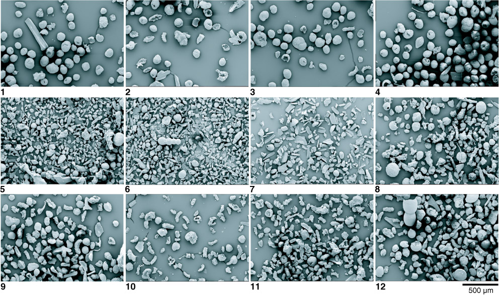
Plate P3. Scanning electron microscope pictures of washed residues selected from the three intervals: (1–4) above the clay-rich layer, (5–8) within, and (9–12) below 1, 2. Sample 198-1209A-23H-3, 108–109 cm. 3, 4. Sample 198-1211B-14H-4, 7–8 cm. 5, 6. Sample 198-1209A-23H-3, 132–133 cm. 7. Sample 198-1211B-14H-4, 32–33 cm. 8. Sample 198-1210A-22H-3, 32–33 cm. 9, 12. Sample 198-1212B-10H-5, 11–12 cm. 10, 11. Sample 198-1209A-23H-4, 7–8 cm.