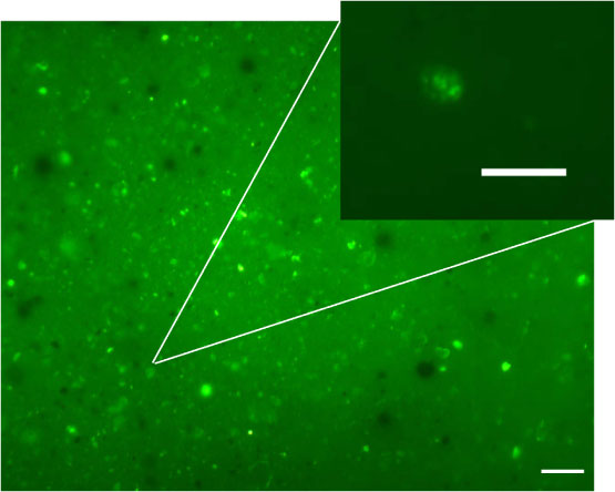
Figure F71. Photomicrograph of sediment showing distinct single small nucleic acids containing objects and bacterial microcolonies after staining with SYBR Green I (Sample 200-1223-1H-1, 0-5 cm). The inset shows a DNA-stained bacterial microcolony at higher magnification. Note the single cells embedded in extracellular polymeric substances. Scale bars for both figures represent 10 µm.