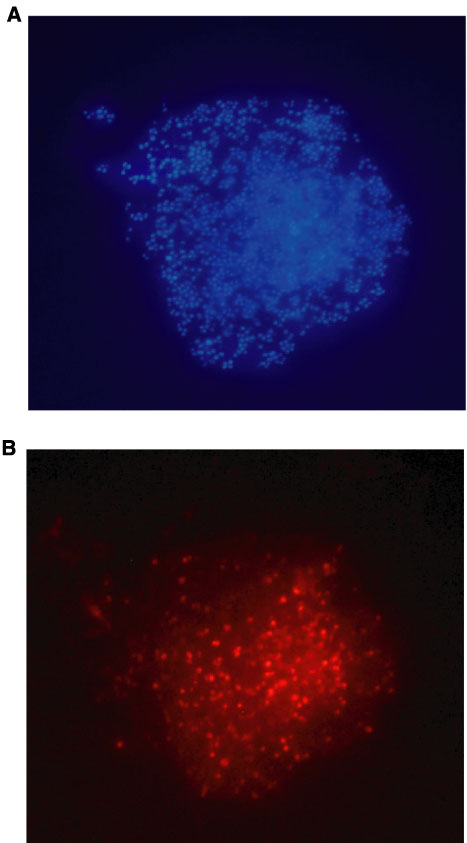
Figure F74. Photomicrograph showing single bacterial cells within bacterial colony CFU16, cultured from altered vitric tuff, after (A) simultaneously staining with the DNA-binding fluorochrome DAPI and (B) hybridization with the CY3-labeled probe SRB385Db, specific for sulfate-reducing bacteria. Note that not all cells given in A did respond to the specific hybridization shown, indicating at least two different bacterial specimens exist within the bacterial colony.