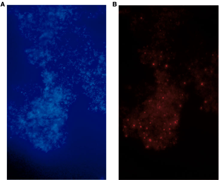
Figure F75. Photomicrograph showing single bacterial cells of bacterial colony CFU1, obtained from altered vitric tuff, after (A) simultaneously staining with the DNA-binding fluorochrome DAPI and (B) hybridization with the CY3-labeled probe LGCa-c, specific for Gram-positive bacteria with low G+C content of DNA.