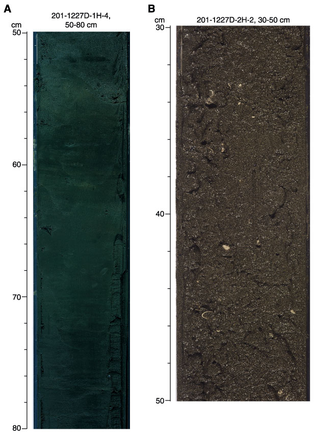
Figure F4.
Close-up photos.
A.
Fine-scale lamination in the diatom ooze of Subunit IA.
B.
Coarse-grained foraminifer ooze.
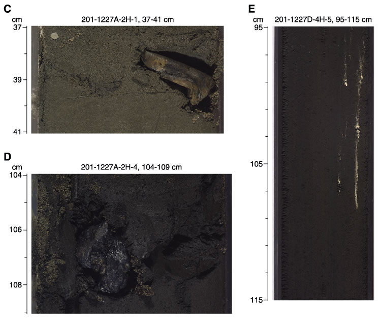
C.
Bone fragment in Subunit IB.
D.
Black massive phosphate nodule from Subunit IIA.
E.
White barite veins.