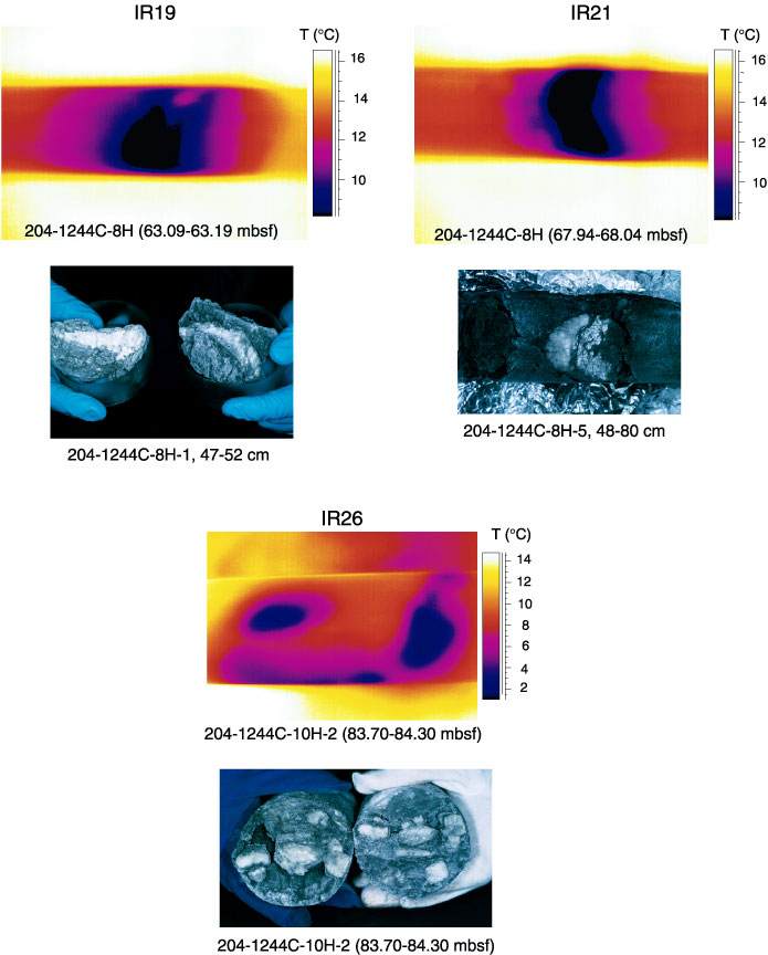
Figure F12. Comparison of IR images and hydrate samples extracted from the core liner. Outer core liner diameter in the IR images is 71.5 mm. Inner core diameter in the photographs is 66 mm. Mismatch between IR anomaly depth and sample depth is because IR scans were taken prior to compressing and cutting core sections.