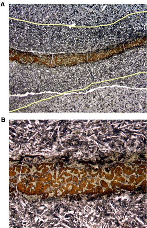
Figure F107. Photomicrograph of Sample 206-1256D-23R-1, 83-85 cm. A. Vein composed of celadonite and iron oxyhydroxides with an adjacent black halo and fresher host rock beyond. Yellow lines mark the boundary between black halo and host rock (field of view = 10 mm; plane polarized light). B. Detail of A showing interstitial celadonite visible in the black halo adjacent to vein (field of view = 2.5 mm; plane polarized light).