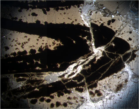
Figure F137.
Photomicrograph showing folded thin bands of flattened and coalesced vesicles within a glassy matrix (Sample
206-1256D-51R-2 [Piece 4, 14-16 cm];
field of view = 0.625 mm; plane polarized light; thin section 151).