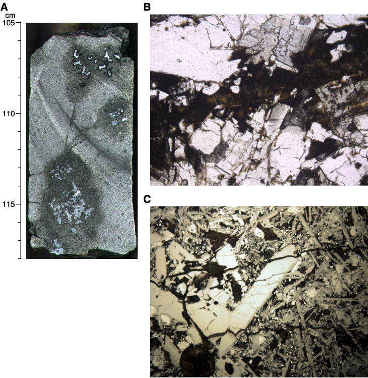
Figure F144. A. Close-up photograph of relationships between veins and amygdules (interval 206-1256D-37R-1 [Piece 8, 106-113 cm]). B. Photomicrograph showing intragranular glassy vein replaced by saponite (Sample 206-1256D-11R-3, 39-42 cm; field of view = 1.25 mm; plane polarized light; thin section 106). C. Photomicrograph showing set of parallel veinlets developed after intragranular microcracks and coated by alteration phases (e.g., saponite). Two veins abut to a vesicle (bottom left) (Sample 206-1256D-46R-1, 64-66 cm; field of view = 1.25 mm; reflected light; thin section 147).