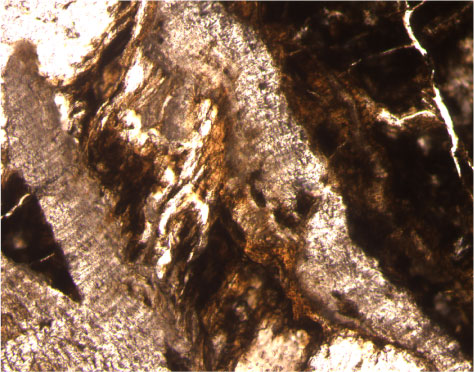
Figure F154. Photomicrograph showing composite ataxial vein filled with saponite, celadonite, iron oxyhydroxides, carbonate, silica, and sulfides (Sample 206-1256C-8R-1 [Piece 5A, 42-47 cm]; field of view = 5 mm; plane polarized light; thin section 20).


![]()