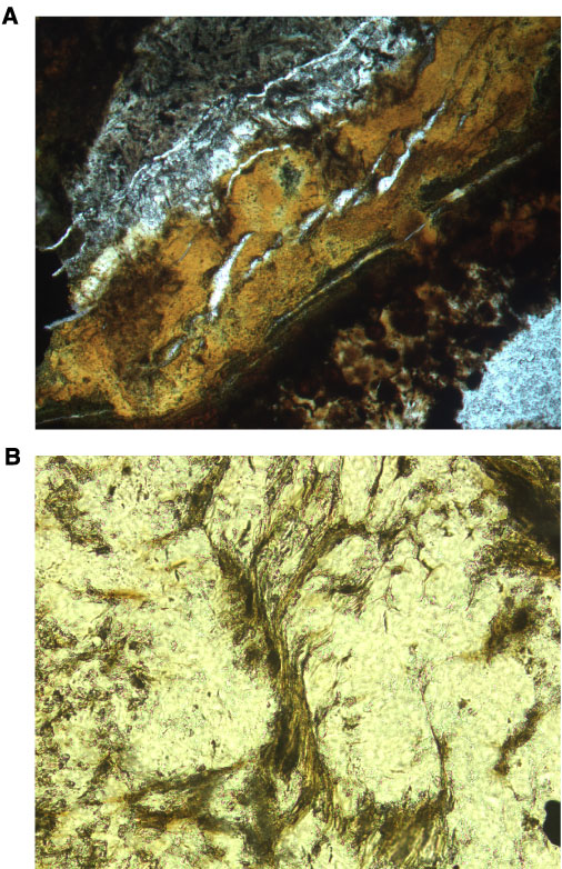
Figure F155. Photomicrographs showing shear deformation in veins. A. Tension gashes opened within iron oxyhydroxide infilling in composite vein (Sample 206-1256C-8R-3 [Piece 13, 136-140 cm]; field of view = 1.25 mm; cross polarized light; thin section 23). B. Sheared saponite fibers (Sample 206-1256C-8R-1 [Piece 5A, 42-47 cm]; field of view = 0.625 mm; plane polarized light; thin section 20).