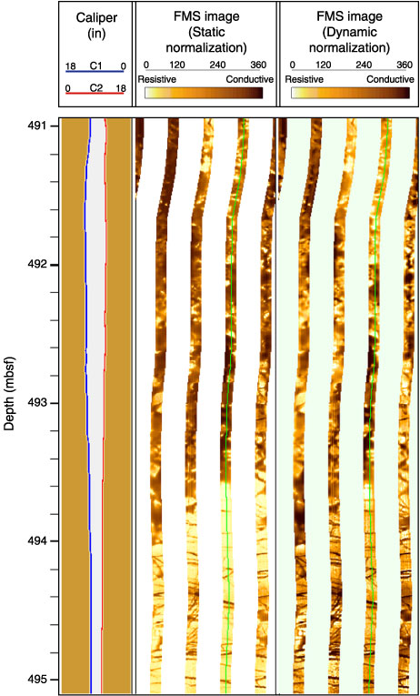
Figure F195. Detailed Formation MicroScanner (FMS) image displaying the transition between a hyaloclastite interval in the upper part of the image (491-493.8 mbsf) and a massive unit in the lower part (493.8-495 mbsf). The hyaloclastites consist of heterogeneous material, with resistive material (basaltic clasts) cemented in a conductive matrix.