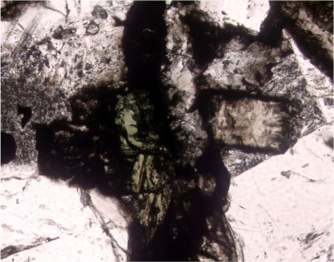
Figure F95.
Photomicrograph showing primary clinopyroxene replaced by secondary green clinopyroxene, adjacent to vein filled with saponite and carbonate (Sample
206-1256C-9R-7, 89-93 cm;
field of view = 2.5 mm; plane polarized light).