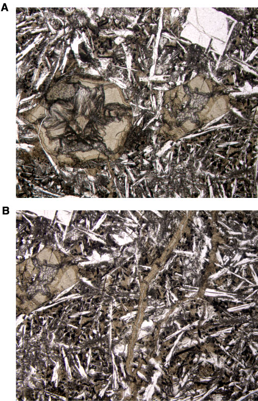
Figure F97. Photomicrograph showing saponite occurrences (Sample 206-1256D-46R-1, 64-66 cm). A. Saponite replacing an olivine phenocryst, filling a vesicle, and within interstitial areas. Image is close to saponite veinlets shown in B (field of view = 2.5 mm; plane polarized light). B. Saponite veinlets (field of view = 2.5 mm; plane polarized light).