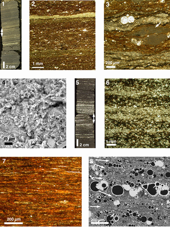
Plate P1. Transmitted light microscope (TLM) photographs (unless stated otherwise). 1. Shipboard Geoscan II image showing laminated pelagic carbonate-rich interval (interval 207-1257B-21R-2, 26–46 cm; 195.06 mbsf; Turonian). Double arrow indicates position of thin section shown in 2 and 3. 2, 3. Sample 207-1257B-21R-2, 33–36 cm (195.13 mbsf; Turonian); (2) micritic white laminae and dispersed planktonic foraminifers of variable size; (3) rare large fecal pellet (arrow). 4. SEM photograph parallel to bedding plane of thin white lamina within predominantly dark colored sediment interval, showing nannofossils concentrated in fecal pellet (Sample 207-1261B-7R-2, 51.5–67 cm; 579.99 mbsf; Coniacian/Santonian). 5. Shipboard Geoscan II image showing laminated planktonic foraminiferal packstone (interval 207-1259A-59R-4, 100–120 cm; 544.83 mbsf; Turonian). Double arrow indicates position of thin section shown in 6. 6. Enlargement of 5 showing variation in concentrations of planktonic foraminifers (Sample 207-1259A-59R-4, 110–113 cm; 544.93 mbsf; Turonian). 7. Example of small fecal pellets and organic matter in organic-rich sediment (Sample 207-1258B-45R-4, 114–117 cm; 401.90 mbsf; OAE2). 8. BSE image of laminated sediment showing potentially seasonal alternation between irregular laminae with planktonic foraminifers separated by thin clay-rich laminae (white arrows) (Sample 207-1259B-23R-3, 114–115.5 cm; 538.14 mbsf; Turonian).