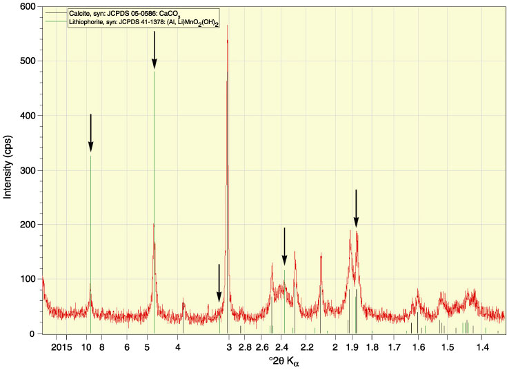
Figure F16.
X-ray diffraction pattern from Sample 208-1264A-26H-5, 25 cm. Arrows highlight the positions of lithiophorite peaks.