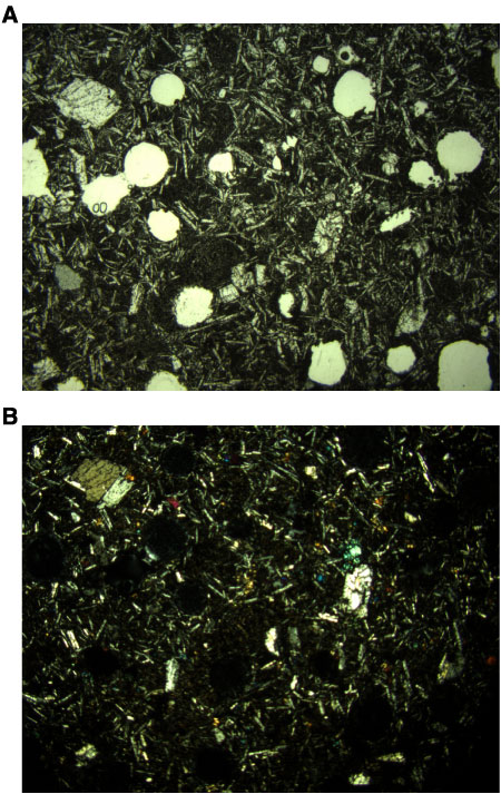
Figure F3.
Photomicrographs showing typical vesicular basalt (Sample
209-1269B-1R-1 [Piece 7, 44–46 cm]
) (blue filter; field of view = 11 mm).
A.
Plane-polarized light; image 1269B_001.
B.
Cross-polarized light; image 1269B_002.