Figure F19.
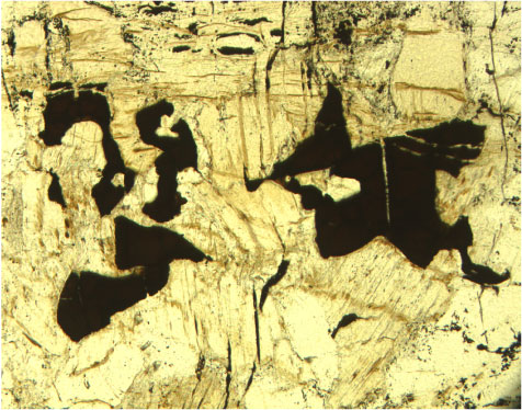
Photomicrograph showing vermicular spinel enclosed in altered orthopyroxene crystals (pale brown) (Sample
209-1270C-1R-1, 72–75 cm
) (cross-polarized light: blue filter; field of view = 1.4 mm; image 1270C_016).