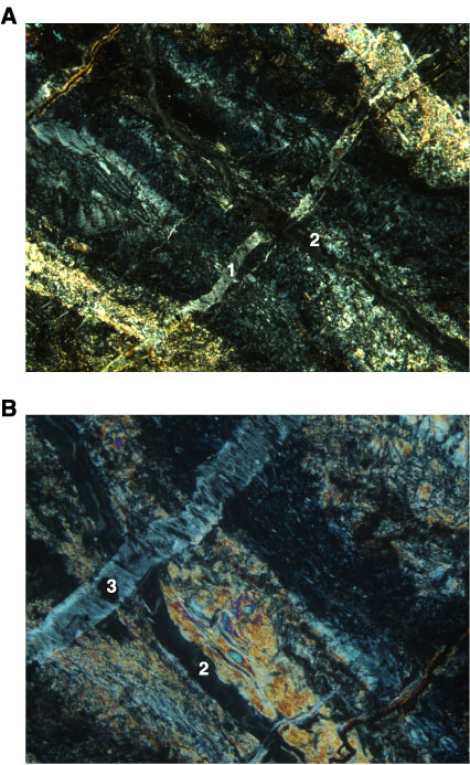
Figure F47. Photomicrographs showing three discrete serpentine vein generations (Sample 209-1270D-6R-1, 18–21 cm). A. Chrysotile vein (1), which crosscuts an altered and deformed gabbroic intrusion, is subsequently cut by a second generation of chrysotile veins propagating along the shear zone (2). An iron oxide vein in the top left corner is also crosscutting the gabbroic intrusion (cross-polarized light: blue filter; field of view 2.75 mm; image 1270D_016). B. This image shows an adjacent part of the same thin section as the one shown in A. Here, a late chrysotile vein (3) crosscuts a second generation serpentine vein (2) in the gabbroic shear zone. The iron oxide vein in the bottom right corner is the same as in A (cross-polarized light: blue + light gray filters; field of view = 1.4 mm; image 1270D_017).