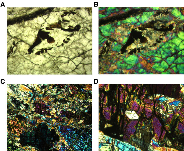
Figure F16. A, B. Photomicrographs showing an alteration phase and spinel enclosed within olivine (Sample 209-1271B-11R-1, 88–91 cm); (A) plane-polarized light: blue + dark gray filters; field of view [FOV] = 0.7 mm; image 1271B_013; (B) cross-polarized light [XPL]: blue + light gray filters; FOV = 0.7 mm; image 1271B_014. C, D. Coarse olivine grains in amphibolite (Sample 209-1271B-13R-1, 20–24 cm) (XPL: blue + light gray filters); (C) FOV = 2.75 mm; image 1271B_100; (D) Close-up view of C; euhedral amphibole enclosed within olivine (FOV = 0.7 mm; image 1271B_099).