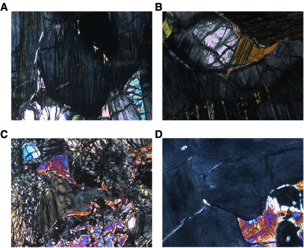
Figure F15. Photomicrographs of orthopyroxene. A. Incipient recrystallization of orthopyroxene into subgrains surrounded by clinopyroxene. A large spinel is included in the orthopyroxene porphyroclast. Clinopyroxene lamellae grew within the orthopyroxene subgrain along the contact with this spinel. In the lower right corner, a small grain of olivine is present together with clinopyroxene at the junction between the two orthopyroxenes and a large olivine crystal (Sample 209-1274A-4R-1, 52–54 cm) (cross-polarized light [XPL]; field of view [FOV] = 1.4 mm; image 1274A_028). B. Kinked and partially recrystallized orthopyroxene. Clinopyroxene mantles a subgrain. Lamellae of clinopyroxene grew within the orthopyroxene along the kink (Sample 209-1274A-7R-1, 101–105 cm) (XPL; FOV = 1.4 mm; image 1274A_047). C. Cluster of two orthopyroxenes, with clinopyroxene developed around them. Note that all the clinopyroxene is in optical continuity (Sample 209-1274A-7R-1, 48–51 cm) (XPL; FOV = 1.4 mm; image 1274A_039). D. Close-up of same sample shown in C showing that the orthopyroxenes are divided into subgrains containing interstitial clinopyroxene (XPL; FOV = 0.7 mm; image 1274A_041).
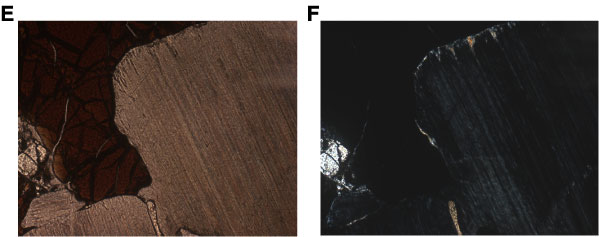
E, F. Contact between a large spinel and orthopyroxene. Thin
lamellae of clinopyroxene grew within orthopyroxene along the contact.
Clinopyroxene also surrounds a small orthopyroxene grain to the left; (E) Sample
209-1274A-11R-1,
6770 cm (plane-polarized light; FOV = 1.4 mm; image 1274A_057);
(F) Sample 209-1274A-11R-1,
6770 cm (XPL; FOV = 1.4 mm, image 1274A_058).