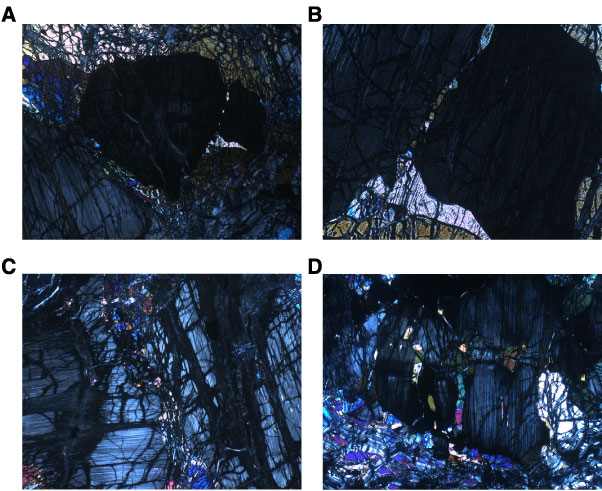
Figure F16. Photomicrographs of orthopyroxene. A-C. Orthopyroxene separated into two grains. The orthopyroxene on the left is separated from the orthopyroxene on the right by a microscopic granular assemblage of olivine, clinopyroxene, and spinel (Sample 209-1274A-3R-1, 92-94 cm) (cross-polarized light [XPL]). (A) Field of view (FOV) = 5.5 mm; image 1274A_025. (B) Close-up showing small grains of olivine and clinopyroxene filling the cracks. A larger clinopyroxene grew at the junction between the two orthopyroxene grains and adjacent olivine (FOV = 1.4 mm; image 1274A_ 026). (C) Close-up, after rotation, of the contact between the two orthopyroxenes in A, showing the fine-grained assemblage of olivine, clinopyroxene, and spinel between them (FOV = 2.75 mm; image 1274A_034). D. Incipient subdivision of kinked orthopyroxene, with microfracture filled with small grains of olivine, clinopyroxene, and spinel (Sample 209-1274A-4R-1, 52–54 cm) (XPL; FOV = 5.5 mm; image 1274A_029).