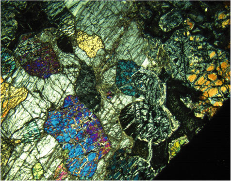
Figure F18. Photomicrograph showing resorbed olivine in orthopyroxene oikocryst. Note how the olivine is isolated by orthopyroxene along the crystal margins (Sample 209-1275B-6R-2, 27-30 cm) (cross-polarized light: blue + dark gray filters; field of view = 11 mm; image 1275B_046).