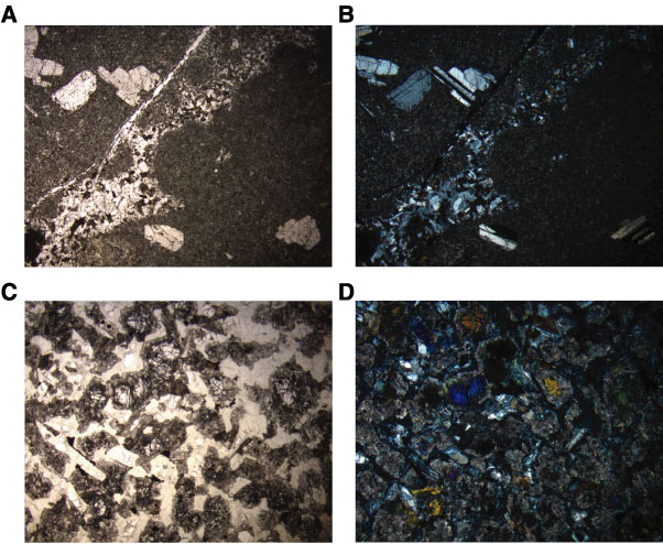
Figure F20. Photomicrographs showing alteration of gabbro. A, B. Granophyre in a porphyritic diabase (Sample 209-1275D-36R-1, 70-72 cm) (field of view = 11 mm); (A) plane-polarized light (PPL): blue + light gray filters; image 1275D_025; (B) cross-polarized light (XPL): blue + light gray filters; image 1275D_026. C, D. General view of altered olivine gabbro (Sample 209-1275D-33R-1, 22-24 cm) (field of view = 11 mm); (C) PPL: blue + light gray filters; image 1275D_023; (D) XPL: blue + light gray filters; image 1275D_024).