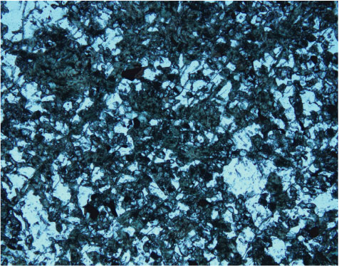
Figure F44.
Photomicrograph showing amphibole veinlets and amphibole-filled microcracks (Sample
209-1275B-17R-1, 60–62 cm
) (plane-polarized light: blue + gray + dark gray filters; field of view = 2.75 mm; image 1275B-021).