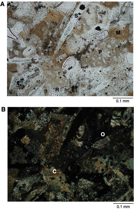
Figure F10. Photomicrographs of grainstone from Unit 1 (Sample 210-1276A-3R-4, 47-49 cm). A. View showing fragments of miliolid (M; tan) and other planktonic foraminifers (P) and siliceous sponge spicules (hollow tubes; S). Porous structure at bottom edge is opaline radiolarian test (R). Black spots are pyrite within porous bioclasts. Very low birefringent chalcedonic quartz fills some pores. B. Same view as A in cross-polarized light to differentiate calcareous (birefringent; C) from opaline (isotropic; O) components.