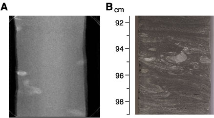
Figure F107. Comparison of CT scan and core photograph from the deformed top of a gravity-flow deposit. A. The white spots on the CT image, not clearly identifiable on the core photograph in B, are interpreted as pyrite concretions. B. Convolute lamination is apparent in the core photograph but is not visible on the CT scan in A (Subunit 5C: interval 210-1276A-89R-6, 91.5-99 cm). This interval is also shown in Figure F94.