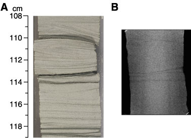
Figure F43. Comparison of core photograph with CT scan. (A) Core photograph of planar- to cross-laminated grainstone compared to (B) CT scan of part of the same interval acquired before core splitting. A and B have the same scale. The well-cemented carbonate grainstone provides little contrast for X-radiography, so only the most prominent clay-rich bands are imaged. This lack of suitable contrast is typical of Units 1-3 (Unit 3: interval 210-1276A-21R-4, 108-119 cm).