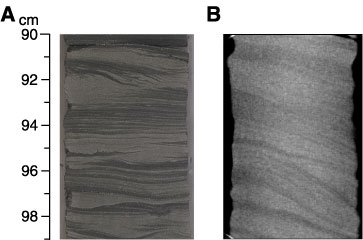
Figure F74. Comparison of sedimentary structures visible on a core photograph compared to a CT scan viewed from a different angle. A. Core photograph. Planar, wavy, and cross lamination. B. CT scan. Cross lamination is visible in the same interval. The photograph and the CT scan show a strike view and a dip view, respectively. The CT scan has the advantage of showing the maximum angle of dip of the bedding that cannot be readily observed in the split cores. This interval is included in Figure F72
(Subunit 5B: interval 210-1276A-45R-2, 90-99 cm).