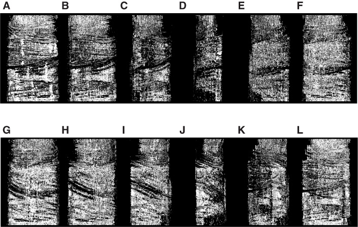
Figure F10.
Three-dimensional X-ray CT images of half-core Sample 210-1276A-44R-5, 27–36 cm, which is the same sample shown in Figure
F1C
and
F1D.
Core image is rotated at 30° steps from A to L.