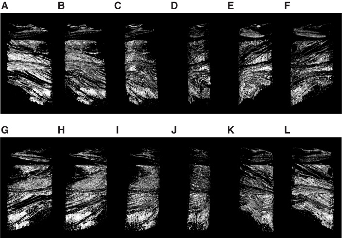
Figure F11.
Three-dimensional X-ray CT images of half-core Sample 210-1276A-48R-3, 56–65 cm, which is the same sample shown in Figure
F2A
and
F2B.
Core image is rotated at 30° steps from A to L.