 Didymocyrtis
avita (Riedel)Didymocyrtis
avita (Riedel)
Didymocyrtis
avita (Riedel)Didymocyrtis
avita (Riedel)Panartus avitus Riedel, 1953, p.808, pl.84, fig.7
Ommatartus avitus (Riedel), Riedel and Sanfilippo, 1971, p.1588, pl.4, fig.6
Didymocyrtis avita (Riedel), Sanfilippo and Riedel, 1980, p.1010
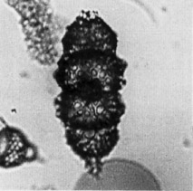
Cortical shell and polar caps of similar general form, kidney-shaped (as in [Didymocyrtis] tetrathalamus (Haeckel)), except that the two polar caps are somewhat narrower than the cortical shell. Pores of the cortical shell subcircular, 3-6 times as broad as the intervening bars, 13-15 on a half equator, 7-9 on the length of a chamber. Walls of the cortical shell bearing large, obtuse protuberances, 1.5-2 times as broad as the pores and half as long, usually concentrated on the widest part of the shell. Pores of the polar caps similar to those of the cortical shell, except for a row of larger pores at the junction with the latter. Surface of the polar caps usually covered with short, acute, conical spines, approximately as long as the diameter of the pores. External medullary shell compressed, its greatest diameter half the equatorial diameter of the cortical shell; the inner medullary shell not clearly visible (modified from Riedel, 1953).
Total length of the shell 162-180 µm; of the apical spines 2-6 µm. Maximum breadth of the cortical shell 80-85 µm; of polar caps 65-75 µm; of the equatorial constriction 70-75 µm; of the external medullary shell 32-35 µm (Riedel, 1953).
D. avita is distinguished from D. penultima [and from all others of the genus] by the absence of spongy columns [and by the heavy protuberances on the cortical shell]. In poorly preserved material, it is often difficult to tell whether specimens are actually columnless, or eroded. Furthermore, the remnants of bars that attached a veil, or a second cap, often resemble the framework of a spongy column. However, if the assemblage is examined as a whole, it can usually be determined whether the species is present.
At the upper end of its range, D. avita loses the tubercles on its cortical shell and evolves to D. tetrathalamus. In some assemblages, broad-based spines on the cortical shell of D. tetrathalamus give the appearance of tubercles (Sanfilippo et al., 1985).
D. avita has a tuberculate, equatorially constricted cortical shell with large caps at each pole, and no spongy columns. The tubercules are prominent in the early part of the range, and become less pronounced with time. Polar caps are sometimes multiple, and a veil may enclose the shell (Sanfilippo et al., 1985).
D. avita is found in all localities of middle Pliocene age from latitudes lower than 40°. It evolved from Didymocyrtis penultima and into Didymocyrtis tetrathalamus within the Spongaster pentas Zone.
D. avita arose from D. penultima and evolved into D. tetrathalamus.
Additional illustrations may be found in Riedel and Sanfilippo, 1971, pl.4, fig.6; 1978b, pl.2, fig.9.
![]()