![]()
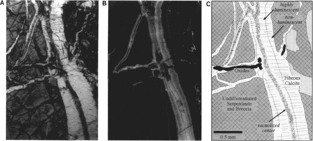
Figure 7. Fibrous veins in Sample 149-899B-16R-2, 69-72 cm, representing a common vein type at both Sites 897 and 899. A. Plane-polarized light photomicrograph showing fibrous calcite veins with distinct vacuolized calcite near their centers; fibers are oriented approximately perpendicular to the vein walls. B. Cathodoluminescence image of same view, showing nonuniform luminescence across the veins; the vacuolized cores are highly luminescent; the adjacent growth displays moderate and relatively uniform luminescence, but shows a sharp transition into nonluminescent calcite at the vein margins. Crosscutting of the luminescent calcite by nonluminescent calcite indicates antitaxial growth of the fibers. C. Interpretive sketch of same view. See Figure 6 caption for explanation of symbols.
![]()