![]() Figures F1-F8.
Figures F1-F8.
![]() Tables
T1-T4.
Tables
T1-T4.
F1. Site locations and generalized surface hydrography.

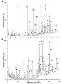
F2. Total ion chromatograms of the total lipid fractions.
F3. Total ion chromatogram of the apolar fraction.

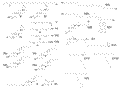
F4. Compound structures of biomarker lipids.
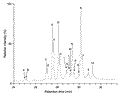
F5. Partial total ion chromatogram of the TLC-5 fraction.
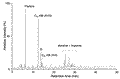
F6. Mass spectrum of compounds from the TLC-6 fraction.
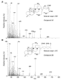
F7. Total ion chromatogram of the apolar fraction of the desulfurized and hydrogenated polar fraction.
F8. Concentrations of quantified biomarker lipids.
T1. Summary of samples analyzed.
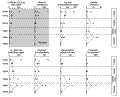
T2. Compounds identified in selected samples.
T3. Steroids identified in selected samples.
T4. Concentrations of quantified biomarker lipids and compound indices.