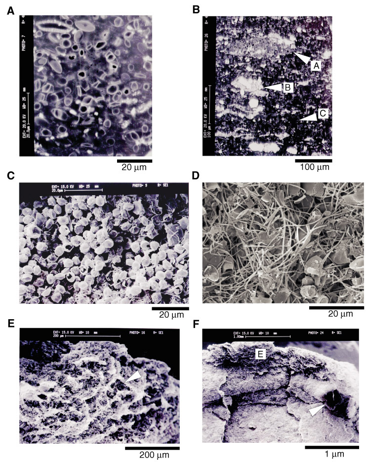
Figure F4. A. BSEI micrograph showing Chaetoceros resting spore-dominated diatom-ooze lamina (Sample 178-1098A-6H-1, 144 cm). B. BSEI micrograph showing the variation of fabric within the diatom-ooze laminae. A = closely packed Chaetoceros resting spores, possible remnants of fecal pellet; B = closely packed terrigenous aggregate, again possible remnants of fecal pellet; C = area of higher porosity, a result of burrows being preserved within the diatom ooze structure (see part E below) (Sample 178-1098A-6H-3, 4.5-8.5 cm). C. SEI micrograph of fractured lamina-parallel surface through diatom ooze. Ooze is comprised almost exclusively of Chaetoceros resting spores with few detached setae (Sample 178-1098A-6H-1, 54.1 cm). D. SEI micrograph of lamina-parallel fracture surface through diatom ooze. Ooze is comprised of Chaetoceros resting spores and ~50% detached setae from the vegetative stage of the Chaetoceros. Both C and D demonstrate the gradation within diatom-ooze laminae from CRS ooze dominated by detached setae in the lower parts to CRS ooze with very few detached setae in the higher parts (Sample 178-1098A-6H-1, 54.4 cm). E. SEI micrograph of vertical fracture surface through a diatom-ooze lamina. Microtunnels and pores are indicated by arrows. These are interpreted as remnant burrow structures (Sample 178-1098A-6H-3, 54-54.3 cm). F. SEI micrograph of vertical fracture surface from a diatom-bearing terrigenous lamina up into a diatom ooze lamina through a bioturbated contact. A large tubelike burrow runs along the contact, one end of which is indicated by an arrow. E = the position of E, above. Below the burrow, you can see the frayed edges of setae sublaminae within the diatom-bearing terrigenous lamina (also see Fig. F5E) (Sample 178-1098A-6H-3, 54-54.3 cm).
![]()