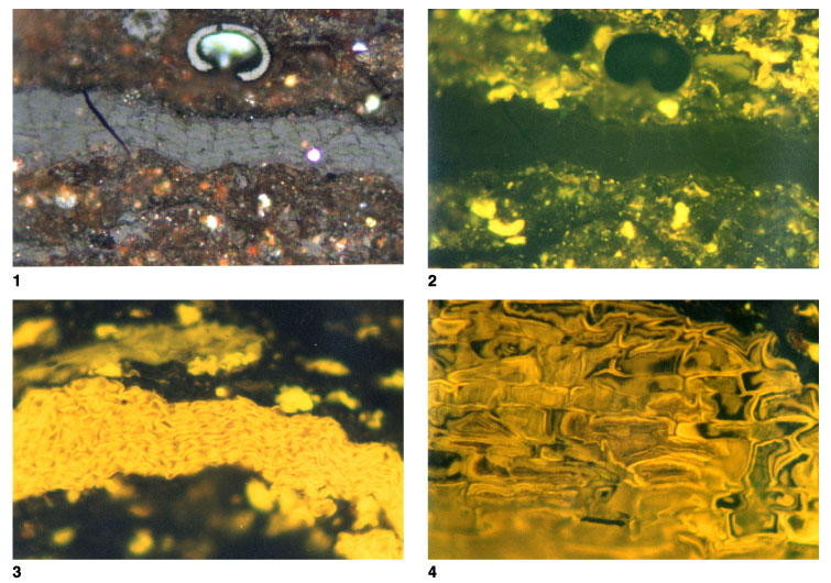
Plate P10.
Coal and shaly coal-cork cells and suberinite. 1.
T5818; Hole 1109D; 969.89 mbsf. Shaly coal with a prominent layer of
telovitrinite showing cork cell structures. The horseshoe-shaped white entity is
a broken fungal teleutospore. Small amounts of pyrite suggest some marine
influence, but this could be postdepositional. Smaller amounts of degraded
telovitrinite and detrovitrinite are present, and abundant liptinite is seen in
the fluorescence-mode plate (reflected light; field width = 0.22 mm;
telovitrinite reflectance = 0.28%, teleutospore reflectance = 0.46%). 2.
T5818; Hole 1109D; 969.89 mbsf. Same as figure 1, but in fluorescence mode. The
outlines of the cell structures within the telovitrinite are weakly shown by
fluorescence from the primary cell walls. Note that fluorescence from these cork
cell tissues is less intense than that from normal woody tissues in many of the
earlier plates. Abundant liptinite is present within the clay-rich layers and
has affinities to resinite and suberinite (reflected light; field width = 0.22
mm; telovitrinite reflectance = 0.28%; teleutospore reflectance = 0.46%). 3.
T5818; Hole 1109D; 969.89 mbsf. Strongly fluorescing layer that appears to
represent suberinite, although in general appearance it is similar to a
sporangium. Polishing hardness is relatively high (reflected light; field width
= 0.22 mm; mean vitrinite reflectance = 0.28% for sample). 4.
T5818; Hole 1109D; 969.89 mbsf. Strongly fluorescing suberinite seen in section
approximately parallel with the surface of the wood. The typical oblong shape of
cork cell is seen, and a compound cell wall structure can be resolved. This
consists of a weakly fluorescing middle lamella flanked by more strongly
fluorescing secondary cell walls. The secondary cell walls are thicker but are
thin compared with the secondary cell walls of some cell types; see, for
example, Plate P8
(reflected light; field width = 0.22 mm; mean vitrinite reflectance = 0.28% for
sample). Click on image or number to see enlargement.