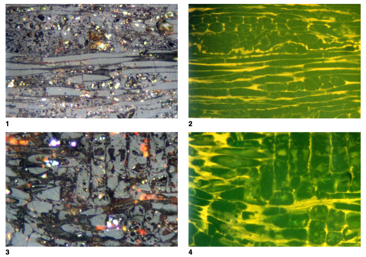
Plate P3.
Vitrinite showing fluorescence from cell walls. 1.
T5809; Hole 1109D; 387.86 mbsf. Longitudinal section of wood preserved as
telovitrinite. The cell lumens are mostly filled with humic material, but the
degree of compressions is small and some parts of the lumens are open. Cell
walls are much lower in reflectance and are strongly fluorescing. Their
reflectances are within the range normally associated with liptinite, but they
do not represent suberinite. Reflectances of the cell walls are lower in the
lower part of the field, but the cell contents have relatively consistent
reflectances across the various tissue types (reflected light; field width =
0.22 mm; vitrinite reflectance [cell contents] = 0.37%, [cell walls] = 0.12%). 2.
T5809; Hole 1109D; 387.86 mbsf. Same as figure 1, but in fluorescence mode.
Longitudinal section of wood with the cells outlined by the fluorescence of the
cell walls. The structures present are probably xylem, seen in longitudinal
section, with some medullary ray tissues in the upper part of the field.
Fluorescence of the cell walls is similar to that for suberinite, but the form
of the cells shows that this material is not from cork cells and cannot strictly
be referred to the maceral suberinite (reflected light; field width = 0.22 mm;
vitrinite reflectance [cell contents] = 0.37%, [cell walls] = 0.12%). 3.
T5809; Hole 1109D; 387.86 mbsf. Oblique section of wood preserved as
telovitrinite. The cell lumens are partially filled with humic material, but
open lumens are still present and little compaction has occurred. Cell walls are
much lower in reflectance and are most easily seen in fluorescence mode. The
vertical cells represent a medullary ray structure (reflected light; field width
= 0.22 mm; vitrinite reflectance [cell contents] = 0.34%). 4.
T5809; Hole 1109D; 387.86 mbsf. Same as figure 3, but in fluorescence mode.
Oblique section of wood preserved as telovitrinite with cell structures outline
by fluorescing cell walls. Some structures are probably present within the cell
walls, and some fluorescence can be seen from the floors of some cells, with the
fluorescence being transmitted through the cell contents (reflected light; field
width = 0.22 mm; vitrinite reflectance [cell contents] = 0.34%). Click on image
or number to see enlargement.