Plate P6.
Suberinite and cutinite. 1.
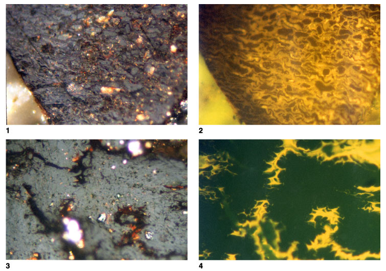
T5843; Hole 1115C; 562.86 mbsf. Telovitrinite and suberinite, with the
suberinite showing positive alteration on prolonged irradiation with the
fluorescence excitation beam. Vitrinite reflectance = 0.32% prior to alteration
and 0.26% after 2 hr of irradiation. The left side of the field was not
irradiated (reflected light; field width = 0.22 mm; vitrinite reflectance [cell
contents] = 0.32%). 2. T5809;
Hole 1109D; 387.86 mbsf. Same as figure 1, but in fluorescence mode.
Fluorescence from the suberinite is much stronger from the area where alteration
has occurred (reflected light; field width = 0.22 mm; vitrinite reflectance
[cell contents] = 0.32%). 3.
T5822; Hole 1114A; 64.72 mbsf. Telovitrinite with cell structure poorly defined
but with cutinite well preserved. The cutinite is difficult to distinguish from
desiccation fractures in white light, but is clearly visible in fluorescence
mode (reflected light; field width = 0.22 mm; mean vitrinite reflectance =
0.33%). 4. T5822; Hole
1114A; 64.72 mbsf. Same as figure 3, but in fluorescence mode. Cutinite probably
seen is section oblique to the surface of the wood. The section angle has
emphasized small reentrants within the outer layers. Fluorescence of the
cutinite is strong, and the form indicates that it is associated with wood
rather than leaf tissues (reflected light; field width = 0.22 mm; mean vitrinite
reflectance = 0.33%). Click on image or number to see enlargement.