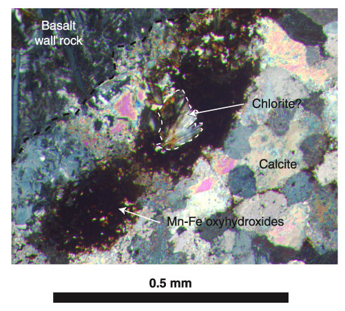
Figure F23. Photomicrograph, with crossed polars, of Sample 187-1157B-3R-1, 97-101 cm (see "Site 1157 Thin Sections"), showing the boundary of a micritic vein (left) that is partially recrystallized to very finely crystalline sparry calcite (right). Note the alteration of wall-rock mineral phases to clay + fibrous amphibole and/or chlorite?. Note also the concentration of Mn-Fe oxyhydroxides + Mn oxides in the micritic part of the vein and their absence from the recrystallized calcite. (See Figure F27 for another view of this section.)
![]()