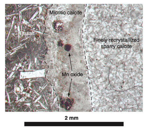
Figure F27. Photomicrograph, with crossed polars, of Sample 187-1157B-3R-1, 97-101 cm (see "Site 1157 Thin Sections"), showing the boundary of a micritic vein (left) partially recrystallized to very finely crystalline sparry calcite (right). Note that Mn oxide is abundant in the micrite but absent from the recrystallized calcite. (Figure F23 shows this sample with crossed polars.)
![]()