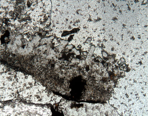
Figure F63. Photomicrograph, at higher magnification, of one of the zeolite "screens" visible in Figure F62 showing the prismatic, longitudinal, and square cross sections characteristic of phillipsite. The zeolite screen is embedded in calcite in Sample 192-1185B-5R-8 (Piece 3, 31-33 cm) (field of view = 1.4 mm; plane-polarized light; photomicrograph ID# 1185B_201).
![]()