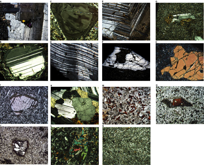
Figure F16. Photomicrographs showing plagioclase phenocrysts. A. Plagioclase megacryst with inclusion of olivine (Sample 197-1205A-15R-2, 138-140 cm) (cross-polarized light; field of view = 5 mm; photomicrograph 1205A-211). B. Rounded plagioclase phenocryst with resorption rim (Sample 197-1205A-13R-3, 27-29 cm) (cross-polarized light; field of view = 1.25 mm; photomicrograph 1205A-206).
C. Compositional zonation in a plagioclase phenocryst from
Subunit 8a (Sample 197-1205A-20R-5,
67-68 cm) (cross-polarized light; field of view = 1.25 mm;
photomicrograph 1205A-216). D. Strain lamellae in a plagioclase
megacryst (Sample 197-1205A-11R-1,
73-75 cm) (cross-polarized light; field of view = 5 mm;
photomicrograph 1205A-204). E, F. Plagioclase phenocrysts from
Subunit 14b (Sample 197-1205A-29R-2,
55-58 cm) (cross-polarized light; field of view = 5 mm): (E) zoned,
fractured, and strained plagioclase phenocryst (photomicrograph 1205A-236); (F)
highly embayed plagioclase phenocryst (photomicrograph 1205A-237). G.
Plagioclase-olivine glomerocrysts (Sample 197-1205A-29R-2,
55-58 cm) (cross-polarized light; field of view = 5 mm;
photomicrograph 1205A-235). H. Highly embayed olivine
phenocryst from Unit 21 (Sample 197-1205A-37R-5,
28-29 cm) (cross-polarized light; field of view = 5 mm;
photomicrograph 1205A-257). I. Partially altered olivine
phenocrysts in Subunit 3b (Sample 197-1205A-8R-1,
59-61 cm) (plane-polarized light; field of view = 1.25 mm;
photomicrograph 1205A-190). J. Partially altered olivine
phenocryst rimmed by biotite in Unit 27 (Sample 197-1205A-43R-2,
58-60 cm) (plane-polarized light; field of view = 5 mm;
photomicrograph 1205A-274). K. Glomerocryst from Subunit 5b,
composed of plagioclase, pyroxene, opaque minerals, and partially altered
olivine (Sample 197-1205A-16R-2,
84-86 cm) (cross-polarized light; field of view = 5 mm;
photomicrograph 1205A-214). L. Subophitic texture in Subunit
3b, with a large groundmass clinopyroxene crystal enclosing a partially altered
olivine phenocryst (Sample 197-1205A-12R-2, 114-116 cm)
(cross-polarized light; field of view = 1.25 mm; photomicrograph 1205A-205). M.
Close-up view of groundmass olivine completely altered to Fe oxyhydroxide or
iddingsite (Sample 197-1205A-24R-2,
124-130 cm) (plane-polarized light; field of view = 1.25 mm;
photomicrograph 1205A-222). N. Strain bands in trachytic
texture, Subunit 3b (Sample 197-1205A-10R-2,
73-75 cm) (cross-polarized light; field of view = 5 mm;
photomicrograph 1205A-202). O. Sheared, relict olivine
phenocryst in Subunit 29b (Sample 197-1205A-44R-2,
110-112 cm) (cross-polarized light; field of view = 1.25 mm;
photomicrograph 1205A-262). Click on letter or image for enlargement.